The Miscellaneous Millinery- Paget's Disease
Anubha Bajaj*
Department of Diagnostics histopathology & Cytopathology Laboratory, India
- *Corresponding Author:
- Anubha Bajaj
Department of Diagnostics histopathology & Cytopathology Laboratory, India
Tel No: 00911141446785
E-mail: anubha.bajaj@gmail.com
Received Date: August 13, 2021; Accepted Date: November 16, 2021; Published Date: November 26, 2021
Citation: Bajaj A (2021) The Miscellaneous Millinery- Paget’s Disease, J Neoplasm, Vol:6 No:2.
Commentary Article
Preface Paget’s disease of bone is achronic, multifactorial skeletal disorder associated with anomalous bone growth, excessive remodelling and comprehensive, diffuse pain within the musculoskeletal system. The commonly discerned disorder manifests classic radiological appearance such as expansive and coarse bony trabecular pattern. Paget’s disease of bone was initially scripted by Sir James Paget in 1877.
The condition is additionally denominated as Paget’s "osteitis deformans" or "osteodystrophica deformans" or "collage of matrix madness" and is accompanied by rapid bone resorption within the osteolytic phase, brisk configuration of bone within the mixed osteoclastic and osteoblastic phase followed by a burnt out, osteo-sclerotic phase associated with an accrual of disorganized bone mass [1-3].
Disease Characteristics Paget’s disease of bone demonstrates excessive osteoclastic activity followed by compensatory elevation of osteoblastic activity. Consequently, weak, disorganized, minimally compact, vascular bone is engendered which is susceptible to fractures.
Generally, Paget’s disease of bone occurs within fourth to fifth decade. Elderly individuals are commonly implicated wherein Paget’s disease involves around 11% of subjects exceeding 60 years. Majority (90%) individuals are above >55 years and the disease is exceptional in individuals beneath < 40 years.
Caucasians are commonly affected whereas the disorder is exceptional in Scandinavians, Chinese, Japanese, Africans and African-Americans. Singular bone is implicated in around one third( 33%) instances. Although the condition can involve singular or multiple bones, axial skeleton such as vertebral column, asymmetric pelvic bones, skull and proximal segment of long bones as the femur are frequently implicated. Polyostotic disease variant is frequently discerned, in contrast to monostotic variant.
Majority (85%) of subjects display polyostotic lesions with incrimination of pelvis, vertebral column and skull. Around 15% individuals delineate asymptomatic, monostotic bone involvement of tibia, ilium, femur, skull, vertebrae or humerus. The condition is exceptional in hands, feet, ribs and fibula.
Paget’s disease of bone is a non contiguous disorder although a pre-existing site can be implicated. The condition depicts a mild male predominance.
The disease commences with osteoclastic activity followed by an admixture of osteoclastic and osteoblastic process. Subsequently, osteoblastic activity is observed which culminates in malignant degeneration.
Disease Pathogenesis Of obscure aetiology, Paget’s disease of bone is contemplated to be a disease of osteoclastic dysfunction. It is posited that osteoclasts generate a unique cytokine confined exclusively within the bone marrow of incriminated subjects designated as interleukin 6.
Paget’s disease delineates enhanced bone resorption with decimated bone mass and osteolytic activity. Osteoblasts engendered from the bone appear on account of a sensory stimulus which elevates osteolytic activity.
Genetic susceptibility, environmental factors or viral infection with paramyxovirus may contribute to disease development. Paget’s disease of bone may be engendered due to slow virus infection of paramyxovirus as the virus may be isolated from the osteoblasts. An association with histocompatibility leucocyte antigen (HLA) is observed. Also, siblings demonstrate an enhanced possibility of disease occurrence.
Clinical Elucidation Majority(75%) of subjects are asymptomatic at initial disease representation. Incriminated subjects manifest with localized pain, tenderness, warmth due to hyperaemic, hyper-vascular lesions, enhanced bone magnitude, bowing deformity, kyphosis of vertebral column, decimated mobility and range of motion.
Additionally, complication related symptoms may be discerned. Paget’s disease of bone demonstrates pyrexia, erythema and persistent warmth upon incriminated hyper-vascular bone leading to high output congestive cardiac failure.
Paget’s disease of bone is a progressive disorder categorized into distinctive stages of disease continuum denominated as
• Early, incipient, destructive stage of Paget’s disease which is comprised of lytic lesions with significant osteoclastic activity.
• Intermediate, active stage is composed of an admixture of osteoblastic and osteoclastic activity.
• Delayed stage is comprised of disease inactivity constituted of commingled sclerotic and blastic lesions.
Subjects may be asymptomatic and majority of instances are discerned by an incidental radiograph. Symptomatic individuals manifest pain within bones and joints, diffuse joint stiffness, significantly enlarged, deformed skull, musculoskeletal deformities, deafness due to implication of petrous temporal bone, migraine, fractures, heart failure, cranial nerve neuropathies, and headache and jaw deformities. Lumbar spine, sacrum and skull are commonly involved.
Generally, pain which worsens upon weight-bearing is observed. Frequently, symptoms are mild and subjects display localized pain due to micro-fractures or nerve compression. Secondary osteoarthritis ensues on account of weakened femur or tibia. Additionally, chalk-like fractures of tibia, fibula or femur are observed [4-6].
Spinal cord injuries occur on account of spinal fractures. Subjects may depict “leontiasis ossea” or a weighty cranium, “platybasia” associated with invagination of base of skull secondary to weakened bone along with compression of posterior fossa.
Paget’s disease of bone is associated with neoplasms such as giant cell tumour, giant cell granuloma or sarcoma which is discerned in around ~5% subjects with severe, polyostotic disease.
Physical examination demonstrates bone deformity, angulation, and localized pain upon palpation and warmth upon disfigured bones. Alterations of gait and balance are discerned.
Incomplete fractures of tibia and fibula are common in Paget’s disease. Minimal injuries can engender a fracture. Fracture of the femur frequently appears within the sub-trochanteric region.
Paget’s disease of the axial skeleton is associated with enhanced cardiac output in around 20% subjects. Additionally, calcified aortic stenosis is discerned. Histological Elucidation Typically, Paget’s disease of bone incriminates bone architecture into distinct phases denominated as osteolytic, mixed and osteosclerotic phase. Aforesaid disease stages may occur simultaneously or asynchronously.
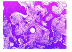
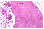
Upon microscopy, enhanced osteoclastic and osteoblastic activity is observed. Acute stage of Paget’s disease of bone primarily demonstrates woven bone along with focal, mosaic configuration of lamellar bone predominantly delineating irregular cement lines. Osteoclasts are preponderantly accumulated upon the bone surface. Osteoclasts appearing within osteolytic phase may depict around ~100 nuclei.
Chronic stage of Paget’s disease of bone is comprised of thickened bony trabeculae and thick bones along with finely delineated fibrosis of bone marrow. Osteolytic phase is associated with foci of bone resorption due to significant quantities of aberrant osteoclasts imbued with several nuclei. Subsequently appearing osteoblastic phase is composed of disorganized, fragmented and irregular bone development. Irregularly constituted bone particles akin to a jigsaw are pathognomonic ofPaget’s disease of bone.
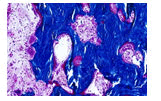
Progressive disease is constituted by a predominant osteoblastic phase with excessive configuration of course, fibrous bone. Bone marrow space is incorporated with vascularized fibrous tissue. Irregular, coarse, disorganized bone configured in Paget’s disease is devoid of centralized vascular articulations or Haversian canals. Configured new bone is inadequately mineralized and lacks structural integrity. Reticulin stain can be employed to highlight disorganized lamellar bone.
Differential Diagnosis Paget’s disease of bone arising within the skull requires a segregation from conditions such as
• hyperostosis frontalis interna demonstrating thickened internal table of frontal bone fibrous dysplasia which usually arises in young adults below< 30 years and commonly incriminates outer table of the calvarium. Polyostotic fibrous dysplasia demonstrates eccentric atrophy of bone cortex.
• Vertebral lesions of Paget’s disease of bone require a distinction from vertebral haemangioma which delineates fine indentations within the bone trabeculae.
Paget’s disease of bone requires a demarcation from conditions such as chronic osteomyelitis, osteomalacia, osteoporosis, osteoarthritis, renal osteodystrophy, osteopenia, bone modifications due to radiation therapy, malignant metamorphosis or secondary deposits from a primary carcinoma and reactive bone arising adjacent to a carcinoma.
Investigative Assay Paget’s disease of bone exhibits elevated serum alkaline phosphatase, hyper-uricaemia and urinary hydroxy-proline along with normal values of serum calcium and phosphate.
Paget’s disease of bone can be appropriately evaluated on bone scan and plain radiographs. The condition denominates an elevation of markers of bone destruction such as N-telopeptide.
Values of hydroxy-proline, C-telopeptide, N-telopeptide and procollagen N terminal peptide require evaluation. Secondary hyperparathyroidism is discerned in nearly 10% instances due to calcium imbalance with increased demand and reduced intake.
Bone scans can satisfactorily ascertain preliminary disease, extent of disease and therapeutic follow up [7-9].
Plain radiographs depict foci of arthritis, fractures and macroscopic bone lesions. Preliminary lesions are radiolucent. Lesions of long standing duration demonstrate enhanced bone density, increased incidence of micro-fractures along with absent demarcation between cortex and medulla although distinction between normal and incriminated bone may be well defined. Florid, extensive disease may extend into circumscribing soft tissue.
Plain radiographs demonstrate specific features contingent to disease stage as
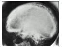
Skull bones in stage I display features of osteoporosis circumscripta with enlarged, well-defined lytic lesions appearing within inner aspect of outer table of the skull along with intact inner table. Admixture of lytic and sclerotic lesions within the skull bones engender a cotton wool appearance. Inner and outer tables of the calvarium are incriminated by diploic widening.
Platybasia and basilar invagination associated with the skull superimposed upon facial bones is denominated as a Tam o' Shanter sign.
• Early stage depicts osteolytic lesions with lucent zones. Subsequently, bone enlargement and coarse bony trabeculae are observed. Sclerotic changes occur eventually. Bone destruction may ensue with malignant transformation.
Paget’s disease of bone arising within the vertebral column frequently manifests with sclerosis and thickening of the bone cortex confined to the vertebral margins with the emergence of picture frame sign, usually associated with mixed phase disease. Lateral radiographs depict a flattening of normal concavity of anterior perimeter of vertebral bodies, denominated as squaring of the vertebrae. Bone trabeculae are coarse and depict vertical thickening.
Pelvic bones display a thickened bone cortex and sclerosis of ilio-pectineal arch and ischio-pubic arch with consequent appearance of pelvic brim sign and obliteration ofKohler’s teardrop. Protrusion of acetabulum is observed along with enlargement of pubic rami and ischium. Modifications of pelvic bones are asymmetric and frequently discerned on the right side.
Long bones depict subchondral radiolucent zones along with V-shaped foci of osteolysis progressing towards the diaphysis, features which are denominated as blade of grass or candle flame sign. Exceptionally, the disease is confined to diaphysis instead of subchondral bone, commonly within the tibia.
Additionally, lateral curvature of femur and anterior curvature of tibia is engendered.
• Magnetic resonance imaging (MRI) demonstrates variable signal intensities contingent to diverse disease stages.
• Dominant signal intensity upon T1 weighted imaging and T2 weighted imaging in Pagetic bone, akin to mature adipose tissue, as frequently encountered in longstanding disease.
• Minimal signal intensity on T1 weighted imaging and enhanced signal intensity on T2 weighted imaging, denominating a “speckled” appearance as encountered with granulation tissue, hyper-vascularity and oedema discerned in early, mixed, active disease
• Minimal signal intensity upon T1 weighted imaging and T2 weighted imaging indicative of compact bone or fibrous tissue as discerned in late, sclerotic stage.
Bone scintigraphy with Technetium -99m (Tc-99m) can be employed to define extent and distribution of disease. Paget’s disease of bone depicts a significant uptake in various disease stages although uptake may be normal in the burnt-out, sclerotic, quiescent phase
Technetium -99m uptake within vertebral bodies and posterior elements configure an inverted triangle as discerned on posterior planar imaging, simulating and denominated as a Mickey Mouse sign or the "heart" sign or "clover" sign or "T" sign or "champagne glass" sign.
Diffuse uptake of technetium-99m within the mandible may articulate a bearded appearance.
Therapeutic Options Paget’s disease of bone appearing in asymptomatic subjects devoid of active disease with abnormal biochemical parameters may not require therapeutic intervention.
Paget’ disease of bone associated with diffuse pain, bone defects incriminating weight bearing bones, deformities of skull and progressive bone alterations necessitate treatment.
Commonly, drugs such as bisphosphonates, calcitonin, supplementation with calcium and vitamin D and painkillers such as acetaminophen or non-steroidal anti-inflammatory drugs (NSAIDs) are prescribed.
Paget’s disease of bone associated with symptoms are appropriately treated with bisphosphonates which can decimate bone turnover, augment healing of osteolytic lesions and alleviate bone pain. Analgesics and non-steroidal anti-inflammatory drugs(NSAIDs) are employed for pain relief.
Paget’s disease of bone progressing into an osteosarcoma can be managed with surgical intervention or palliative options such as amputation of incriminated limb. Younger individuals can be offered limb salvage surgery along with en bloc tumour resection with a wide tumour perimeter. Pain associated with pathological fractures requires radiation and internal fixation.
Chemotherapy is non-effective in treating sarcoma appearing in Paget’s disease of bone. Revision surgery is frequently indicated.
Cauda equina syndrome and nerve compression associated with Paget’s disease of bone can be treated with laminotomy.
Paget’s disease of bone may be associated with osseous weakening with deformities, pathological fractures, enhanced possibility of osteoarthritis, sensi-neural or conductive deafness, compression of vestibulocochlear nerve within the internal auditory canal, reduced bone mineral density of the cochlear capsule, fixation of middle ear ossicles, neural compression, cranial nerve paresis, basilar invagination, hydrocephalus and brainstem compression along with emergence of secondary neoplasms such as osteosarcoma or giant cell tumour of the skull. Additionally, high output congestive cardiac failure, left to right shunts and decreased peripheral resistance is observed.
Paget’s disease of bone exhibits complications such as secondary osteoarthritis, vertigo, deafness, dental malocclusion, tinnitus, cauda equina syndrome, Hashimoto’s thyroiditis, osteopetrosis or Dupuytren’s contracture.
Hyperparathyroidism and extramedullary haematopoiesis may ensue. Paget’s disease of bone may terminate in a lethal Paget’s disease associated sarcoma.
Exceptionally, a fatal osteosarcoma appears which demonstrates rapidly progressive bone swelling or bone pain. Giant cell tumour may arise in Paget’s disease of bone and preponderantly incriminate facial bones. Vertebral fractures are associated with acute spinal cord compression. Appropriately treated preliminary Paget’s disease of bone is accompanied by a favourable prognosis. Progression of Paget’s disease of bone can be curtailed although a comprehensive cure is unachievable. Polyostotic disease is associated with adverse outcomes, in contrast to monostotic disease.
Morbidity in Paget’s disease of bone arises due to pathological fractures, chronic pain, bone deformities and neurological complication. Degeneration and metamorphosis into a sarcoma exemplifies a significantly inferior prognosis.

Open Access Journals
- Aquaculture & Veterinary Science
- Chemistry & Chemical Sciences
- Clinical Sciences
- Engineering
- General Science
- Genetics & Molecular Biology
- Health Care & Nursing
- Immunology & Microbiology
- Materials Science
- Mathematics & Physics
- Medical Sciences
- Neurology & Psychiatry
- Oncology & Cancer Science
- Pharmaceutical Sciences
