Peculiarities of neoplasms , appeared after total body irradiation and homeostasis parameters in rats.
Elisaveta Snezhkova1*, Sakhno LA1, Bardakhivska KI1, Nikolaev VG1, Voronina OK2, Zadvornyi TV 3, Todor IN3, Lukianova NY3, Chekhun VF3 and Melnyk VO4
1Department of Means and Methods of Adsorptive Therapy, R.E. Kavetsky Institute of Ex perimental Pathology, Oncology an d Radiobiology, National Academy of Sciences of Ukraine, 03022 Kyiv, Ukraine
2Department of Cytology, Histology and Reproductive Medicine, Institute of Bi ology and Medicine, Taras Shevchenko National University of Kyiv, 01033 Kyiv, Ukraine
3Department of Monitoring of Tumor Process and Therapy Design, R.E. Kavetsky Institute of Experimental Pathology, Oncology and Radiobiology, N ational Academy of Sciences of Ukraine, 03022 Kyiv, Ukraine
4Department of Biochemistry, National Cancer Institute of Ukraine, 03022 Kyiv, Ukraine
- *Corresponding Author:
- Elisaveta Snezhkova
Department of Means and Methods of Adsorptive Therapy, R. E. Kavetsky Institute of Experimental Pathology,
Oncology and Radiobiology,
National Academy of Sciences of Ukraine, 03022 Kyiv,
Ukraine,
Tel: 380953124221;
Email: lisasne@hotmail.com
Received: February 18, 2022, Manuscript No. IPJN-22-11398; Editor assigned: February 21, 2022, PreQC No. IPJN-22-11398 (PQ); Reviewed: March 07, 2022, QC No. IPJN-22-11398; Revised: March 11, 2022, Manuscript No. IPJN-22-11398 (R); Published: March 18, 2022, Invoice No. IPJN-22-11398
Citation: Snezhkova E, Sakhno LA, Bardakhivska KI, Nikolaev VG, Voronina OK, et al. (2022) Peculiarities of Neoplasms Appeared After Total Body Irradiation and Homeostasis Parameters in Rats. J Neoplasm Vol:7 No:1
Abstract
Tissue damage and disruption of metabolic processes as a result of Total Body Irradiation (TBI) could lead to tumorigenesis. About a year after TBI (12 ± 1 mth) 76% of female rats have visually detected tumors, which were histologically classified as benign fibro adenomas. Metabolic disorders and hematological changes observed during this period indicate disturbances in the homeostasis system. The blood lactate level in all tumor-bearing animals was statistically higher than in control (healthy) rats-a typical phenomenon for neoplasm growth. In the majority of animals with neoplasms (63%) the tumor weight and the levels of lactate and Lactate Dehydrogenase (LDH) in the blood were in direct relation (coefficient=tumor weight/ lactate level>0.5<1.5) and histological analysis revealed the signs of biphasic hyperplasia of glandular lobes and connective tissue stroma, associated with secretory and proliferative activities in tumor. In the conditional grouping of animals with relatively high values of coefficient (≥ 1.5), neoplasms were represented by fibrous and glandular tissues presenting a predominant stromal fibrous component, associated with the prevalence of high proliferation in tumor. While in the rat conditional grouping (20% of animals) with relatively low coefficient (<0.5) an epithelial structure with homogeneous basophilic content predominated in the glandular lumens suggesting the domination of high secretory activity in tumor.Thus, not only the elevated blood lactate or LDH level but also the ratio of tumor mass to this lactate or LDH value seems to be an informative indicator of the peculiarities of tumor process.
Keywords
Rat; Total body irradiation; Tumorigenesis; Ratio of tumor mass to lactate; LDH
Introduction
The risk of neoplasms appearance after Total Body Irradiation (TBI) in moderate doses has been demonstrated in numerous experimental investigations [1,2] as well as in the clinical studies of the Japanese atomic bomb survivors and of the Chernobyl accident consequences [3]. Tumors have also been reported in patients undergoing radiation treatment and diagnostics [4,5]. The appearance of tumors after irradiation is often related to the fact that the radiation can lead to life-threatening multiple organ failure, dysfunction syndrome and inflammation [6,7]. After TBI, a variety of diseases frequently develop, such as; extrahepatic bile obstruction, intrahepatic cholestasis, infiltrative liver disease, which is accompanied by increased serum Alkaline Phosphatase (ALP) [8] and also pancreatitis with an elevated level of amylase [9]. The most classically recognized symptom of acute radiation sickness is hematopoietic syndrome resulting in reduced count of platelet, leukocytes, and erythrocytes. Even low levels of exposure can lead to bone marrow failure, potentially lethal hemorrhage, or infections [10]. Decreased partial arterial oxygen pressure and an increased level of carboxyl groups are well known signs of the pathological states in acute radiation syndrome and tumorigenesis [11-12].
Tumor hypoxia leads to therapeutic resistance [11,13] and also promotes aggressive tumor behavior and metastases. Tumor clones dramatically alter their metabolic activity to meet the metabolic demands of relentless cell division. The highly conserved metabolic pathway of fermentative glycolysis is exploited by rapidly growing tissues and tumors [14]. Lactate generation is a cellular process necessary for maintaining glycolytic flux and facilitating the removal of pyruvate from the cell. The interconversion of pyruvate to lactate is mediated by Lactate Dehydrogenase (LDH) and results in the oxidation of NADH to NAD+. The role of lactate in cancer was described by Otto Warburg in 1927, prominently produced by glycolytic cells within tumors and a major fuel for oxidative cancer cells, and therefore a diagnostic agent [15-18].
Lactate exchange across membranes is gated by Monocarboxylate Transporters (MCTs) which are up regulated in tumors. Thus, elevated lactate levels [15-17], up-regulation of LDH [18], and the expression of monocarboxylate transporter-MCTs [19] are prognostic of tumor progression. A highly glycolytic tumor metabolism is also associated with resistance to conventional therapies [20]. The parameters of glycolytic tumor metabolism are important predictors of tumor progression and patient’s survival.
In the present study we aimed to investigate the main hematological and biochemical homeostasis parameters and peculiarities of tumorigenesis in rats after total body irradiation in sub-lethal doses.
Materials and Methods
Animal model
Two weeks before the start of the study (approved by IEPOR Committee on Bioethics, protocol No 4 dated 16 April 2015), thirty two white, random bred, female rats weighing 140 g from IEPOR animal house, were taken for adaptation period and had ad libitum access to standard food and water in accordance with the provisions of the European Convention for the Protection of Vertebrate Animals Used for Experimental and Other Scientific Purposes (Strasbourg, 1986). At the beginning of experiment all animals were randomly allocated into groups:
• Irradiated rats (n=25)
• Healthy rats (n=7)
Weighed and examined every month for visual tumor appearance. On month 12 ± 1in major part of irradiated animal’s tumor was detected visually. This period was selected as optimal for animal’s sacrifice because of the risk of tumor necrosis appearance unfavorable for histological study. After dissection of all 32 animals, tumors were found in 19 rats after irradiation and this group was named Ir and Tum (n=19), the group of irradiated animal without tumor Ir (n=6) and healthy rats-control (n=7).
Irradiation
Ionizing radiation was delivered to 8-week-old rats weighing 194 g ± 18 (prior to irradiation), using an X-ray RUM-17 irradiator with a working current of 10 mA, 0.5 mm Cu filter, 30 cm target distance in rotating box with 4 separate section (24 × 15 cm) for 4 rats, exposure time was 11 min. Control group animals underwent the same procedure, excluding irradiation. The absorbed sub-lethal dosage per rat was 6 Gy (54.5 cGy/min during 11 min), which was applied in the morning between 10-11 am.
Blood collection for blood cell count, biochemical parameters, lactate, po2, pco2 , ph
Terminal citrate blood samples and heparin blood samples (for lactate, PO2 , PCO2 , PH) were obtained via punctue of abdominal vein.
Blood cell count
Blood cell counts were performed using light microscope.
Lactate, PO2, PCO2 , PH level in blood: actate, PO2 , PCO2 , PH levels were measured using gas analyzer ABL800-Flex (Radiometer, Denmark, Copenhagen)
Blood biochemical parameters
All biochemical parameters of serum were measured using reagent kits of Beckman Coulter Corporation and auto-analyzer (Beckman Coulter AU-480, USA). Manufacturer’s instructions were followed.
Methods for determining: the Alkaline Phosphatase (ALP) [21]; Alanine Aminotransferase (ALT), Aspartate Aminotransferase (AST) [22]; Amylase [23], Lactate Dehydrogenase (LDH) [24]; Ca2+ [25]; Creatinine [26]; Glucose [27]; Phosphorus [28]; Urea [29]; Uric acid [30]. Total protein was measured by the biuret method.
Histology of tumor tissues
Tumor tissues were carefully isolated for histological examination, weighed, measured and fixed in 4% neutral buffered formalin. Fixed tissues were dehydrated and embedded in paraffin and then cut into serial 4 μm thick sections. The histopathological characteristics were obtained on tumor tissue sections after hematoxylin and eosin staining in 19 rats with tumor.
Statistical analysis
Mean values and standard deviations were calculated. Differences between groups were evaluated statistically with Fisher then with Student’s t-test for independent samples. Group differences were considered significant when p<0.05. For biochemical and hematological parameters statistical analysis of the results was carried out using Mann-Whitney nonparametric comparison. Statistical significance was set at<0.05.
Results
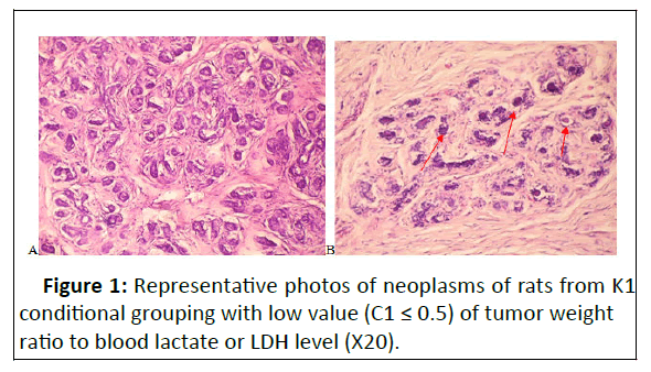
Twelve months after irradiation in 76% of females (19 from 25, group-Ir and tum) tumors was detected visually in the lower part of the animal's body associated with mammalian glands. Histological analysis indicates that all the tumors found were fibro of the breast (Figures 1-3). Fibroadenoma is a benign neoplasm, in which the lobular structure of the gland is preserved. But the proliferation of both glandular and stromal elements is clearly manifested, atypia is not observed in any of the components. Glandular and stromal components of the gland are clearly visible on histological preparations of tumors.
The glandular parenchyma is represented by secretory (tubuloalveolar) units and their clusters with the layers between formed by fibrous connective tissue. No tumor was found in any control.
The statistical increase of alkaline phosphatase was observed and compared with the control group. Amylase activity in the plasma of rats in both irradiated groups was statistically higher one year after irradiation than in the control (Table 1).
| Rat’s group |
ALP, U/L |
ALT, U/L |
Amylase, U/L |
AST, U/L |
LDH, U/L |
Ca2+ mmol/L |
Creatinine, µmol/L |
Glucose, mmol/L |
Phosphorus, mmol/L |
Total protein ,g/L |
Urea, mmol/L | Uric Acid, µmol/L |
|---|---|---|---|---|---|---|---|---|---|---|---|---|
| Control n=7 | 111 ± 33 | 54 ± 17 | 1701 ± 251 | 183 ± 20 | 935 ± 317 | 2.2 ± 0.1 | 41 ± 8.6 | 3.9 ± 0.9 | 2.3 ± 0.7 | 63 ± 8 | 7.1 ± 1.2 | 72 ± 9 |
| Ir n=6 | 250 ± 40* | 66 ± 11 | 2609 ± 380* | 181 ± 33 | 600 ± 59 | 2.4 ± 0.1 | 34 ± 2 | 5.3 ± 0.4 | 1.5 ± 0.1 | 63 ± 3.1 | 5.6 ± 1 | 60 ± 5 |
| Ir&Tum, n=19 | 151± 77 | 57 ± 29 | 2180 ± 679* | 171 ± 84 | 768 ± 336 | 2.0 ± 0.4 | 32 ± 5.5 | 4.0 ± 1.1 | 1.3 ± 0.4 | 55 ± 10.4 | 6.6 ± 1.1 | 78 ± 46 |
| *According to Mann-Whitney test the result is significant at p<0.05 (in comparison with control) | ||||||||||||
Table 1: Alkaline Phosphatase (ALP), Alanine Aminotransferase (ALT), Amylase, Aspartate Aminotransferase(AST), Lactate Dehydrogenase (LDH), Ca 2+, Crea inine, Glucose, Phosphorus, Urea, Total Protein, Uric Acid level in blood plasma of rat’s groups: healthy animals (control), Irradiated (Ir), with tumor detected visually one year after total body irradia ion (Ir and Tum).
± Standard devia ion
| Rat's group | Hb, g/L | WBC, 109/L | RBC,10 12/ L | Eosinophils, 109/L |
Neutrophiles Stab., 10 9/L |
Neutrophiles segmented, 109/L |
Lymphocytes, 109/L |
Monocytes, 109/L | Platelets, 10 9/L |
|---|---|---|---|---|---|---|---|---|---|
| Control, n=7 | 131 ± 12.4 | 10 ± 1.1 | 5 ± 0.9 | 0.04 ± 0.04 | 0.08 ± 0.03 | 2 ± 0.5 | 7 ± 1.6 | 0.5 ± 0.2 | 267 ± 29 |
| Ir, n=6 | 132 ± 1.2 | 9 ± 1.6 | 5 ± 0.2 | 0.04 ± 0.04 | 0.0 ± 0.0 | 2 ± 0.2 | 7 ± 1.5 | 0.2 ± 0.1 | 304 ± 11 |
| Ir&Tum, n=17 | 125 ± 13 | 5.9 ± 1.7*# | 4.5 ± 0.5 | 0.02 ± 0.04 | 0.1 ± 0.03 | 1.2 ± 0.5 | 4.3 ± 1.1 | 0.2 ± 0.2 | 204 ± 57*# |
| *According to Mann-Whitney test the result is significant at p<0.05 (in comparison with control). ≠ According to Mann-Whitney test the result is significant at p< 0.05 (in comparison with Ir) |
|||||||||
Table 2: Hematological parameters of rats (control-healthy animals, irradiated-Ir, with visually detected tumor after irradiation Ir and Tum): Hemoglobin (Hb), White Blood Cells (WBC), Red Blood Cells (RBC), neutrophils stab and segmented, Lymphocytes, Monocytes, and platelets.
± Standard deviation
Hematological parameters in major cases demonstrate the worsening in both irradiated groups. White blood cells and platelets level was found to be significantly fewer in the blood of irradiated animals with a tumor in comparison not only with healthy control but with irradiated animals without tumors (Table 2)
The lactate level was signi icantly elevated in the tumor group compared to the control. In both irradiated groups PO2 was signi icantly reduced, and in the irradiated without tumor one PCO2 increased versus the control group (Table 3).
| Rat’s group | Lactate, mmol/L |
PO2, mm Hg |
PCO2, mm Hg |
PH |
|---|---|---|---|---|
| Control, ( n=7) | 5,3 ± 0,8 | 60.7± 2,3 | 33,8 ± 8,9 | 7,3 ± 0,05 |
| Ir, (n=6) | 6,3 ± 1,2 | 45,6 ± 3,7* | 42,6 ± 1,7* | 7,3 ± 0,05 |
| Ir&Tum, ( n=19) | 6,6 ± 1,9* | 41,1 ± 6,2* | 38,3 ± 5,9 | 7,3 ± 0,1 |
| *According to Student test the result is significant at p<0.05 (in comparison with control) | ||||
Table 3: Lactate, PO2, PCO2, and pH in the blood of rat’s groups: healthy animals (control), irradiated ( Ir), with tumor appearance one year after total body irradiation (Ir and Tum).
The first tumor was detected visually five months postirradiation (Table 4) but the majority ones of tumors appeared later in the tenth (n=9) and thirteenth (n=5) month after Irradiation (Table 4).
| № rat |
Month of tumor detection after irradiation |
Tumor Volume, cm3 |
Tumor Weight,g |
LDH, U/L |
Lactate level, mmol/L |
C1 | C2 | K groupings |
|---|---|---|---|---|---|---|---|---|
| 1 | 13 | 0,88 | 2,28 | 1307 | 5,7 | 0,4 | 1,7 | K1 |
| 2 | 10 | 1,05 | 1,8 | 668 | 4,2 | 0,4 | 2,7 | K1 |
| 3 | 11 | 3,3 | 4,24 | 1338 | 8,7 | 0,5 | 3,2 | K1 |
| 4 | 10 | 4,25 | 4,61 | 1370 | 9,4 | 0,5 | 3,4 | K1 |
| 5 | 5 | 2,68 | 2,76 | 1401 | 4,5 | 0,6 | 2,0 | K2 |
| 6 | 10 | 4,5 | 6,66 | 546 | 8,1 | 0,8 | 12,2 | K2 |
| 7 | 13 | 8,77 | 7,7 | 720 | 8,9 | 0,9 | 10,7 | K2 |
| 8 | 9 | 6,21 | 7,82 | 621 | 8,5 | 0,9 | 12,6 | K2 |
| 9 | 10 | 1,98 | 3,41 | 480 | 3,7 | 0,9 | 7,1 | K2 |
| 10 | 10 | 6,63 | 5,62 | 484 | 5,9 | 1,0 | 11,6 | K2 |
| 11 | 13 | 1,98 | 4,76 | 735 | 4,8 | 1,0 | 6,5 | K2 |
| 12 | 10 | 4,35 | 8,32 | 590 | 8,1 | 1,0 | 14,1 | K2 |
| 13 | 10 | 2,77 | 4,75 | 536 | 4,5 | 1,1 | 8,9 | K2 |
| 14 | 13 | 10,58 | 6,85 | 597 | 5,3 | 1,3 | 11,5 | K2 |
| 15 | 13 | 6,84 | 8,2 | 826 | 6,1 | 1,3 | 9,9 | K2 |
| 16 | 10 | 7,6 | 8,71 | 991 | 6,3 | 1,4 | 8,8 | K2 |
| 17 | 8 | 1,79 | 6,63 | 453 | 4,3 | 1,5 | 14,6 | K3 |
| 18 | 10 | 26,8 | 19,46 | 659 | 9,6 | 2,0 | 29,5 | K3 |
| 19 | 8 | 25,3 | 36,8 | 279 | 8 | 4,6 | 131,9 | K3 |
Table 4: Weight and volume of tumors in Ir and Tum group of rats (n=19) and month of their visual detection a ter total body irradiation; blood Lactate Dehydrogenase (LDH) and Lactate levels; C1-coe icient=Tumor weight/Lactate level; C2- coe icient=Tumor weight/ LDH *1000, K-classi ied groupings of animals, where: K1 is the grouping of rats with C1 ≤ 0.5; K2-with C1>0.5<1.5; K3 with C1 ≤ 1.5
The certain correlation of tumor weight (but not tumor volume, measured manually) with lactate and LDH values in the blood of irradiated animals is demonstrated in table 4. The animals were distributed in three groupings named K1-K3, according to the coefficient C1 reflecting the ratio of tumor weight to blood lactate level. In the majority of animals (63.2%, n=12, grouping K2) tumor weight is in direct relation with blood lactate and LDH levels and the C1 coefficient is approximately equal to 1 (>0.5<1.5). In the remaining 36.8% of animals the
lactate and LDH levels are not in direct relation with tumor weight. In grouping of rats classi ied as K1 (n=4) C1 has the value ≤ 0.5 and in the K3 grouping -C1 ≥ 1.5 (n=3, 15.8% of animals). C1 coe icient correlates (except rat № 5 with C1 value 0.6 on the border of K2 and K1 values) with the C2 coe icient (ratio of tumor weight to LDH level) that con irms the correct distribution of animals in these conditional K groupings and the possibility to use blood lactate or LDH levels for classi ication.
We investigated the histological peculiarities of neoplasms in 19 rats of group Ir and Tum, classified and grouped conditionally according to the values of the coefficient C1.
Neoplasms in classified as K1 grouping (n=4) with a relatively low tumor weight ratio to lactate level in blood (C1 ≤ 0.5) produce a tumor comprised of fibrous and glandular tissues with a predominant epithelial component of a clear lobular structure breast (Figure 1, A-B). Glandular structures are formed by a twolayer epithelium without signs of atypia. The inner glandular layer consists of cuboidal cells with normochromic nuclei. Benign processes are recognized by the presence of myofibroblasts. Basophilic content is noticeable in the lumen of many glands and duct and the cystic extension of the ducts can be observed (Figure 1B). The stroma is homogeneous, hypo vascular, with signs of fibrosis. The fusiform fibroblasts with elongated nuclei are seen between collagen fibers. No signs of inflammation and hemorrhages in the tumor are observed in the connective tissue stroma.

Figure 1:Representative photos of neoplasms of rats from K1 conditional grouping with low value (C1 ≤ 0.5) of tumor weight ratio to blood lactate or LDH level (X20).
A. Proliferation of acinar structures consisting of epithelium and myoepithelium surrounded by the basement membrane. Acinuses round and oval, with interlobular fibrous connective tissue, dilatation of the ducts. There are no signs of malignancy.
B. In the lumens of the glands noticeable basophili c homogeneous content (red arrows).
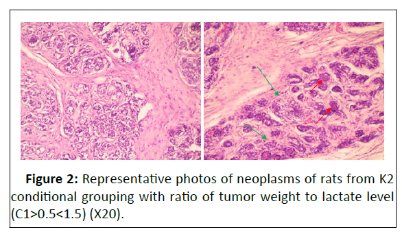
Histological examination of tumors in rats of conditional K2 grouping (n=12) with a tumor weight to blood lactate level ratio C1>0.5<1.5 demonstrated signs of biphasic hyperplasia of glandular lobes and connective tissue stroma (Figure 2 А, В). Neoplasms are represented by hyperplastic breast tissue with a clear lobular structure.

Figure 2:Representative photos of neoplasms of rats from K2 conditional grouping with ratio of tumor weight to lactate level (C1>0.5<1.5) (X20).
Acini are lined with epithelial cells having vacuolated cytoplasm, indicating active synthesis as well as secretion. Epitheliocytes are forming a multilayered epithelium, not typical to the glands but without signs of atypia. Nuclei are of typical size with eu-and hetero-chromatin.
Mitotic figures are not found. In most animals of this group small cystic cavities filled with basophilic homogeneous mass (Figure 2В) are observed. The stroma is represented by welldeveloped collagen fibers, with clearly seen nuclei of fibroblasts between them. The blood vessels in the tumor are dilated and filled with blood cells. Outside vessels, erythrocytes are visible in a stroma as well as in a parenchyma (Figure 2В).
А. Lobular hyperplasia. Round or oval acinuses, between which there are well-developed layers of connective tissue, stromal fibrosis.
В. Proliferation of both glandular and stromal elements. Basophilic homogeneous content is visible in the lumens of the glands (red arrows)
Inflammatory infiltrate in the stroma and glands (green arrows).
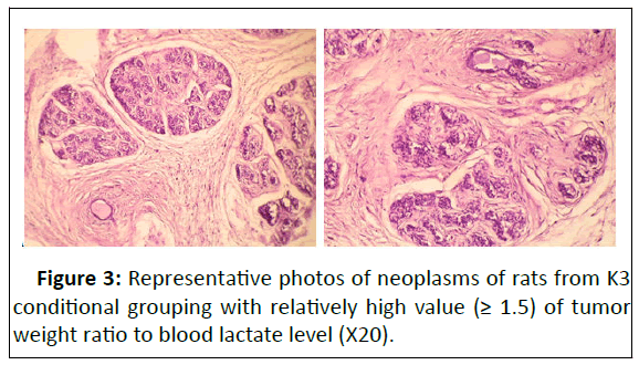
The parenchyma of tumors in rats from K3 conditional grouping (n=3) having relatively high values of ratio tumor weight to blood lactate level (C1 ≥ 1.5 ) is represented by a complex branched tubular-alveolar system, separated from each other by stromal connective tissue. Moreover, the stromal fibrous component predominates in slides (Figure 3, А-B), with clearly seen well-developed fibers and elongated fibroblasts.

Figure 3:Representative photos of neoplasms of rats from K3 conditional grouping with relatively high value (≥ 1.5) of tumor weight ratio to blood lactate level (X20).
А. Вreast fibroadenomas with connective tissue fibrosis. Collagen and bland spindle shaped stromal cells with ovoid or elongated nuclei. Vessels without pathological changes, lined with a single layer of endothelium.
В. Lobular hyperplasia. Densely located small alveoli formed by two-layer epithelium. Well-developed connective tissue stroma around the glands.
In K1, K2, K3 conditional groupings of Ir+Tum group of rats, the histological features of neoplasms are similar in general but had some specific differences. In all groups, the tumors were identified as benign (non-cancerous) breast fibroadenomas with signs of proliferation of epithelial and stromal elements.
In grouping classified as K3 with maximal values of tumor weight ratio to lactate (C1 ≥ 1.5) the connective tissue stroma, was associated with increased proliferation. While in grouping classified as K1, with minimal C1 (≤ 0.5), tumor tissue dominated well developed ductal epithelium with lobular hyperplasia, indicating high secretory activity. In rats of К1 grouping as well as in the most ones of K2 grouping homogeneous basophilic content in the lobule was presented. It indicates the high secretory activity of epithelial cells.
Signs of inflammation were found only in some animals of K2 and K3 groupings. In several rats of K2 grouping histological signs of extensive vascular blood supply in tumors were discovered.
Discussion
In 5-13 months after total body irradiation, 76% of irradiated female rats (Table 4) were found to have benign tumors fibro adenomas, associated with mammalian glands (Figure 1-3). The peak of tumors appearance (79%) occurred 10-13 months after irradiation. No tumor was detected visually in the controls. In our experiment only benign tumors were detected and histologically confirmed, while Bespalov and coauthors [1] reported about 40% of malignant tumors from total number of radiation induced tumors (80% of animals) 16 months after total body gamma-ray irradiation in dose of 4 Gy (1.34 Gy per min). Perhaps this is due to the different doses and sources of rat’s irradiation.
Any way benign tumors as well as malignant ones are progressing and this process is associated with high energetic demands. Lactate and LDH, the main actors of fermentative glycolysis, meet the demand in energy of tumors. The fact of elevation of lactate and LDH level in presence of tumor was described in numerous investigations, [15-18,31,32]. Furthermore, increased blood LDH and lactate values were observed in both malignant [33] and benign [31,32] growth. In our study blood, lactate level in the group of animals with tumor (Ir and Tum) is statistically higher than in healthy animals (Table 3). But the lactate dehydrogenase activity in the same group of animals statistically no, possibly due to high values of the standard deviation (Table 1).
We have tried to find the correlation between the ratio of the mass of tumors to lactate/LDH levels in the blood and the histological peculiarities of tumor tissue. For this, the 19 animals with irradiation induced tumors were divided into three conditional groupings (K1-K3) according to the value (C1) of tumor weight ratio to lactate level (Table 4). We considered it important that the C2 coefficient, which reflects the ratio of tumor weight to blood LDH level (one of the main participants in lactate metabolism), correlates with the C1 coefficient the ratio of tumor weight to lactate level. This means that the same animals can be formed into the three conditional K1-K3 groupings focusing on the values of tumor weight ratio to either lactate or lactate dehydrogenase level in blood.
In the majority of animals with a tumor in K2 grouping (63.20%, n=12) with the value of tumor weight ratio to blood lactate level (C1>0.5<1.5) histological examination demonstrated the signs of biphasic hyperplasia of glandular lobes and connective tissue stroma (Figure 2 А,В), indicating the secretory and proliferative activities of tumor tissues. Neoplasms in K3 grouping (Table 4) with relatively high values of tumor weight ratio to lactate level (C1 ≥ 1.5, n=3) are represented by fibrous and glandular tissues with domination of the connective tissue stromal component (Figure 3, A-B). That shows the prevalence of proliferative activity of tumor cells. In the K1 grouping with relatively low (C1 ≤ 0.5) value of tumor weight ratio to blood lactate or LDH level (Figure1) the epithelial structure with homogeneous basophilic content dominates the glandular lumens. It is associated with prevalence of high secretory activity of tumor. Thus, the values of the ratio of tumor weight to blood lactate or LDH level (C1 and C2 coefficients) correlate with histological specificity of tumor tissues. This makes it possible to evaluate indirectly the peculiarities of tumor state, indicating the prevalence of proliferative activity, as in K3 grouping, or secretory one, as in K1 grouping, or the certain balance between proliferative and secretory activities of tumor cells (K2 grouping).
The metabolic and hematological misbalances that has arisen after irradiation confirms the violation of homeostasis that conducts to tumorigenesis. In the group of irradiated rats without tumors, a significant increase in serum Alkaline Phosphatase (ALP) indicates a metabolic disorder, disrupting homeostasis (Table 1). Markedly elevated ALP is seen predominantly with a number of specific pathologies, such as malignant biliary obstruction, primary biliary cirrhosis, primary sclerosing cholangitis, hepatic lymphoma and sarcoidosis [34].
Amylase activity is depicted in Table 1. A significant increase in the activity of serum amylase (but only 1.5-1.3 fold) is evident in both groups of rats one year after irradiation. Increased activity of amylase and lipase is usually a diagnostic sign of pancreatitis [35]; these enzymes are released from the pancreas into circulation early in the inflammation process. Blood leucocytes and platelets count (Table 2) is statistically lower in the Ir+Tum group indicating an inhibitory effect of the tumor on hematopoiesis of these cells of blood. The statistical drop of PO2 was found in both groups of irradiated animals (Table 3) that indicates the presence of pathological processes. Elevated lactate levels in blood of animals with irradiation-induced tumors (Ir and Tum group), being a sign of the metabolic adaption of tumor cells but is also a component utilized by a variety of inflammatory immune cells [15]. Release of lactate from tumor cells is accompanied by acidification in the tumor microenvironment favoring tumor promotion, angiogenesis, metastasis, tumor resistance [17]. The microenvironment of a tumor consists of a dynamic and complex network of cytokines, growth factors, and metabolic products. These contribute to significant alterations in cell growth, tissue architecture, immune cell phenotype and function. LDH, one of the main actors of lactate metabolism, is released from cells in response to their damage, causing its baseline level to rise in the extracellular space and the bloodstream or other body fluids as well. Because an elevated LDH level was found to be an unfavorable indicator for survival in cancer patients, it was suggested that lactate dehydrogenase can be used as a marker of tumor aggressiveness [33].
The preliminary results obtained in this experiment permit to suggest that not only the elevated level of lactate or LDH in blood but also the ratio of tumor mass, detected by modern methods in clinic, to values of blood lactate or LDH level, may be informative for characterization of the tumor process.
Conclusion
Total body irradiation in sub-lethal doses promoted the alterations of hematological and biochemical parameters of homeostasis in rats and provoked the benign tumors appearance one year after. The ratio value of tumor mass to blood lactate or lactate dehydrogenase level reflects the histological peculiarities of tumors, indicating in this way the state of tumor process.
References
- Vladimir G Bespalov, Valery A Alexandrov, Alexandr L Semenov, Elena G Kovan’ko, Sergey D Ivanov, et al. (2017) The inhibitory effect of meadowsweet (Filipendula ulmaria) on radiation-induced carcinogenesis in rats. Int J Radiat Biol 93:394-401
[Crossref] [Google Scholar] [PubMed]
- Hollander CF, Zurcher c, Broerse jj (2003) Tumorigenesis in high-dose total body irradiated rhesus monkeys--a life span study. Toxicol Pathol 31:209-213
[Crossref] [Google Scholar] [PubMed]
- Thomas G A, Symonds P (2016) Radiation Exposure and Health Effects – is it Time to Reassess the Real Consequences?. Clin Oncol 28(4):231–236
[Crossref] [Google Scholar] [PubMed]
- Gilbert ES (2009) Ionising radiation and cancer risks: what have welearned from epidemiology. Int J Radiat Biol 85:467-482
[Crossref] [Google Scholar] [PubMed]
- Scott Baker k, Wendy M Leisenring, Pamela J Goodman, Ralph P Ermoian, Mary E Flowers, et al. (2019) Total body irradiation dose and risk of subsequent neoplasms following allogeneic hematopoietic cell transplantation Blood 133: 2790–2799
[Crossref] [Google Scholar] [PubMed]
- Kiang JG, Olabisi AO (2019) Radiation: A poly-traumatic hit leading to multi-organ injury. Cell Biosci 1–15
[Crossref] [Google Scholar] [PubMed]
- Nakamura NA (2020) hypothesis: radiation carcinogenesis may result from tissue injuries and subsequent recovery processes which can act astumor promoters and lead to an earlier onset of cancer. Br J Radiol 93: 20190843
[Crossref] [Google Scholar] [PubMed]
- McIntyre N, Rosalki S (1991) Biochemical investigations in the management of liver disease. In: McIntyre R, editor Oxford Textbook of Clinical Hepatology Oxford, England: Oxford University Press. Gynecol Obstet Invest 127:293–309
- Lotfi SA, El-Kabany H ( 2012) Therapeutic Response of Black Tea Extract on Maintenance Pancreas and Intestine of Gamma-irradiated Rats. J Rad Res Appl Sci 5:619 – 632
- Dainiak N (2002)Hematologic consequences of exposure to ionizing radiation. Exp Hematol 30:513–528
[Crossref] [Google Scholar] [PubMed]
- Bertout JA, Patel SA, Simon MC (2008) The impact of O2 availability on human cancer Nature reviews 8:967–975
[Crossref] [Google Scholar] [PubMed]
- Vaupel P (2008) Hypoxia and aggressive tumor phenotype: implications for therapy and prognosis. Oncologist 3:21–26.
[Crossref] [Google Scholar] [PubMed]
- Siemann DW, Hill RP (1983) Quantitative changes in the arterial blood gases of mice following localized irradiation of the lungs. Radiat Res 93:560-566
[Crossref] [Google Scholar] [PubMed]
- Scott K Parks, Wolfgang Mueller-Klieser, Jacques Pouysségur (2020) Lactate and Acidity in the Cancer Microenvironment. Annu Rev Cancer Biol 4:141-158
- Cathal Harmon, Cliona O Farrelly, Mark W Robinson (2020) The Immune Consequences of Lactate in the Tumor Microenvironment. Adv Exp Med Biol 1259:113-124
[Crossref] [Google Scholar] [PubMed]
- Karen G de la Cruz-López,Leonardo Josué Castro-Muñoz, Diego O Reyes-Hernández,Alejandro García-Carrancá, Joaquín Manzo-Merino ( 2019)Lactate in the Regulation of Tumor Microenvironment and Therapeutic Approaches. Front Oncol 9: 1143
[Crossref] [Google Scholar] [PubMed]
- Ricardo Pérez-Tomás, Isabel Pérez-Guillén (2020) Lactate in the Tumor Microenvironment: An Essential Molecule in Cancer Progression and Treatment Cancers 12:324-354
[Crossref] [Google Scholar] [PubMed]
- Sandra Van Wilpe, Rutger Koornstra, Martijn Den Brok, Jan Willem De Groot, Christian Blank et al. (2020) Lactate dehydrogenase: a marker of diminished antitumor immunity. OncoImmunology 9:354-488
- Valéry L Payen, EricaMina, Vincent F Van Hée, Paolo E Porporato, Pierre Sonveaux (2020) Monocarboxylate transporters in cancer. Mol Metab 33:48-6620
[Crossref] [Google Scholor] [PubMed]
- Liu Y, He C, Huang X (2017) Metformin partially reverses the carboplatin-resistance in NSCLC by inhibiting glucose metabolism. Oncotarget 8:75206-75216
[Crossref] [Google Scholar] [PubMed]
- Keiding R, Hörder M, Gerhardt Denmark W, Pitkänen E, Tenhunen R, et al. (1974) Recommended Methods for the Determination of Four Enzymes in Blood. Scand J Clin Lab Invest 33:291-306
- Gerhard Schumann, Roberto Bonora, Ferruccio Ceriotti, Georges Férard, Carlo A Ferrero, et al. (2002 )International Federation of Clinical Chemistry and Laboratory Medicine IFCC primary reference procedures for the measurement of catalytic activity concentrations of enzymes at 37 degrees C. International Federation of Clinical Chemistry and Laboratory Medicine Part 4 Reference procedure for the measurement of catalytic concentration of alanine aminotransferase Part 5 Reference procedure for the measurement of catalytic concentration of aspartate aminotransferase. Clin Chem Lab Med 40:718-733
- AY Foo, R Bais (1998) Amylase measurement with 2-chloro-4-nitrophenyl maltotrioside as substrate. Clin Chim Act 272:137-147
[Crossref] [Google Scholar] [PubMed]
- Amador E, Dorfman LE, Wacker WE (1963) Serum lactic dehydrogenase activity: an analytical assessment of current assays. Clin Chem 9:391-399
[Crossref] [Google Scholar] [PubMed]
- Michaylova V, Ilkova P, (1971) Photometric determination of micro amounts of calcium with arsenazo III. Analytica Chimica Acta, 53:194-198
- Jaffe M (1886) Ueber den Niederschlag welchen Pikrinsäure in normalen Harn erzeugt und über eine neue reaction des Kreatinins. Z Physiol Chem 10:391–400
- Czok R, Barthelmai W (1962) Enzymatische Bestimmungen der Glucose in Blut, Liquor und Harn. Klin Wschr 40:585-589
- Daly JA, Ertingshausen G (1972) Direct method for determining inorganic phosphate in serum with the "CentrifiChem". Clin Chem 18:263-265
[Crossref] [Google Scholar] [PubMed]
- Talke H, Schubert GE (1965) Enzymatic urea determination in the blood and serum in the warburg optical test, Klinische Wochenschrift 43: 174-175
[Crossref] [Google Scholar] [PubMed]
- Fossati P, Prencipe L, Berti G (1980) Use of 3,5-dichloro-2-hydroxybenzenesulfonic acid/4-aminophenazone chromogenic system in direct enzymic assay of uric acid in serum and urine. Clin Chem 26:227-231
[Crossref] [Google Scholar] [PubMed]
- Michael I Koukourakis, Emmanuel Kontomanolis, Alexandra Giatromanolaki, Efthimios Sivridis, Vassilios Liberis (2009) Serum and tissue LDH levels in patients with breast/gynaecological cancer and benign diseases. Gynecol Obstet Invest 19:162-168
[Crossref] [Google Scholar] [PubMed]
- Schwartz MK (1991) Lactic dehydrogenase An old enzyme reborn as a cancer marker. Am J Clin Pathol 96:441–443
[Crossref] [Google Scholar] [PubMed]
- Forkasiewicz A, Dorociak M, Stach K, et al. (2020) The usefulness of lactate dehydrogenase measurements in current oncological practice. Cell Mol Biol Lett 123:25-35
[Crossref] [Google Scholar] [PubMed]
- Allison L, Brichacek, Candice M Brown (2012) Alkaline Phosphatase: A Potential Biomarker for Stroke and Implications for Treatment. Metab Brain Dis 34: 3–19
[Crossref] [Google Scholar] [PubMed]
- Lotfi SA, H El-Kabany (2012) Therapeutic Response of Black Tea Extract on Maintenance Pancreas and Intestine of Gamma-irradiated Rats. J Rad Res Appl Sci 5:619–632

Open Access Journals
- Aquaculture & Veterinary Science
- Chemistry & Chemical Sciences
- Clinical Sciences
- Engineering
- General Science
- Genetics & Molecular Biology
- Health Care & Nursing
- Immunology & Microbiology
- Materials Science
- Mathematics & Physics
- Medical Sciences
- Neurology & Psychiatry
- Oncology & Cancer Science
- Pharmaceutical Sciences
