Primary Ectomesenchymoma of the Testis, Case Report and Review of the Literature
Cruz Medina Cynthia Shanat
Department of Oncology Pediatric, University of Benemérita Universidad Autónoma de Puebla, Puebla, Mexico
Published Date: 2023-10-23DOI10.36648/2576-3903.8.4.46
Cruz Medina Cynthia Shanat*, Ramos Balderas Cesar, Macias Díaz Salvador, Castro Sanchez Andrea, Escamilla Lopez Miriam Ixel, Tellez Bernal Eduardo, Ortega Solar José María, Garrido Hernandez Miguel Angel and Reynier Castañeda Daniel
Department of Oncology Pediatric, University of Benemérita Universidad Autónoma de Puebla, Puebla, Mexico
- *Corresponding Author:
- Cruz Medina Cynthia Shanat
Department of Oncology Pediatric,
University of Benemérita Universidad Autónoma de Puebla, Puebla,
Mexico,
E-mail: ped_hope@hotmail.com
Received date: September 22, 2023, Manuscript No. IPJN-23-17923; Editor assigned date: September 25, 2023, PreQC No. IPJN-23-17923 (PQ); Reviewed date: October 09, 2023, QC No. IPJN-23-17923; Revised date: October 16, 2023, Manuscript No. IPJN-23-17923 (R); Published date: October 23, 2023, DOI: 10.36648/2576-3903.8.4.46
Citation: Shanat CMC, Cesar RB, Salvador MD, Andrea CS, Ixel ELM, et al. (2023) Primary Ectomesenchymoma of the Testis, Case Report and Review of the Literature. J Neoplasm Vol:8 No.4:46.
Abstract
Malignant Ectomesenchymoma (MEM) is an exceptionally rare neoplasm. We present the case of a patient presenting in the testicle, localized disease, with complete surgical resection, treated with high-risk chemotherapy for the rhabdomyosarcoma component. There is a little information in the bibliography about ectomesenchymoma, in Mexico there are no statistics about this pathology, the study of this pathology will give more information about the pathophysiological behavior and the response to treatment and survival of patients.
Keywords
Neural crest; Rhabdomyosarcoma; Mesenchyme; Neuroectoderm; Ectomesenchymoma; Testis
Introduction
Malignant Ectomesenchymoma (MEM) is an exceptionally rare neoplasm. It has been postulated that MEM originates from remnants of the neural crest, called ectomesenchyma [1,2]. Considering the increased migration of neural crest cells during gestation, the different sites of MEM presentation are explained, although the pelvis and perineum are the most affected areas (45.5%), head and neck in second place (20.5%), intracranial in third place (15.9%), extremities in 9% [3]. However, some cytogenetic studies recent molecular studies have revealed that MEM could represent a variant of Rhabdomyosarcoma (RMS) [3,4]. Most MEM present before 5 years of age. It has a slightly higher incidence in men, with a radius of 1.6, 80% are diagnosed before 10 years of age [5].
Histologically speaking MEM has elements of both mesenchyme and neuroectoderm, the mesenchyme component is frequently RMS, particularly of the embryonic subtype, while the neuroectoderm derivative is frequently neuroblastic tumor with various degrees of differentiation from; neoplasm poor in Schwannian stroma to neoplasm rich in Schwannian Stroma with primitive neuroblastic cells to mature ganglion cells. Only in rare cases is there a neuroectodermal component of the Primitive Neuroectodermal Tumor (PNET) type [6,7]. The classification of soft tissue tumors according to the World Health Organization (WHO) identifies as an essential criterion for the diagnosis of MEM the presence of histological component of embryonic RMS, mixed areas with neuroblastic component (neurons, ganglioneuroma, ganglioneuroblastoma or neuroblastoma), confirmed with immunohistochemistry; positivity for desmin, myogenin and synaptophysin [8,9]. The best treatment protocol is not yet defined, particularly the role of chemotherapy and whether it should be focused on the mesenchymal or neuroectoderm component.
Case Presentation
This paper describes the case of a 14-year-old male with a 2- month evolution clinical picture characterized by right testicular pain and growth.
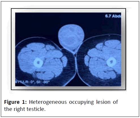
The patient denies a genetic burden for oncological processes, trauma to the genital area, weight loss, fever, lymph node growths or pain in other places. Who are sent to the oncology unit at our institution. On physical examination, normal tegument coloration, adequate hydration, absence of right testicle in scrotal sac, with left testicle in proper location and shape (Figure 1).
Elevation of tumor markers upon arrival of DHL 176 u/l, AFP 1.87 ng/ml and HGC 0.100 mui/ml (requested as a testicular tumor approach). Given the diagnostic suspicion, a tomography of the skull, thorax and abdomen was performed, showing lung parenchyma without alterations, mesenteric lymph node calcifications and the rest of the other structures without alterations.
Results and Discussion
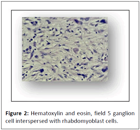
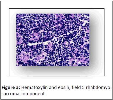
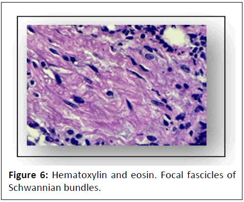
Review of laboratory slides in our institution with a report that corresponds to a malignant neoplasm of mixed strain of the ectomesenchymoma type that is poorly delimited, not encapsulated, architecturally organized into long fascicles, alternating with areas with loose stroma and microcystic changes, focally there are hypercellular basophilic islands and compact within a fibrillar stroma, the cells are spindle-shaped, with eosinophilic cytoplasm, poorly defined cell membrane, pleomorphic nucleus, some with an undulating or serpentine pattern with a hyperchromatic nucleus and others with an enlarged nucleus, with an irregular membrane, vesicular chromatins and an evident nucleus (Figures 2 and 3).
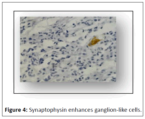
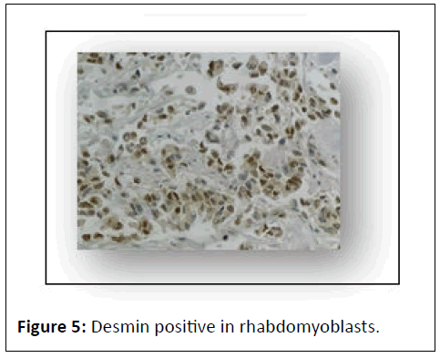
Scattered among the fascicular cells are others with abundant clear eosinophilic cytoplasm, an eccentric nucleus with scattered chromatin and an evident nucleus, surrounded by flat cells with scant cytoplasm and a hyperchromatic nucleus, compatible with ganglion and satellite cells. The basophilic islands are composed of cells with a primitive appearance with little cytoplasm, a hyperchromatic nucleus, with an incospicuous nucleus. The cells are positive for diffuse desmin in the cytoplasm of neoplastic cells, diffuse positive for myogenin in the nucleus of neoplastic cells, focal positive for synaptophysin in the cytoplasm of neoplastic cells (Figures 4-6).
Staged according to TNM. T1b/N0/M0, so it was decided to start treatment with systemic chemotherapy based on vincristine, ifosfamide and actinomycin (via therapy), protocol for sarcomas.
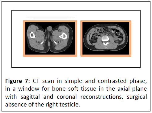
Currently in follow-up and surveillance with the last control of biochemical markers with AFP 0.13 ng/ml and DHL 98 mui/ml. CT scan with absence of right testicle (Figure 7).
Literature review
MEM is a rare sarcoma, it is part of the family of soft tissue sarcomas with a biphenotypic behavior, with co-expression of mesenchymal and ectodermal malignant tissue, there are few cases reported in the literature, in the study by Heng-Chung Kung, et al. to the; published in the Journal of Pediatrics and Neonatology [5] report a review from 2000 to 2021 in which there are only 44 cases reported in the literature; 56.8% presented embryonal rhabdomyosarcoma component, 34.1% presented neuroblastoma component.
Ectomesenchymomas are often confused with rhabdomyosarcomas due to the fact that their neuronal components are easily missed. However, the high plasma concentration of Neuropeptide-Y-as well as immunoreactivity (P-NPY-LI) in ectomesenchymoma can distinguish this type of tumor from rhabdomyosarcoma, which has normal levels of P-NPY-LI [10].
The treatment of MEM is multimodal; surgical resection of the tumor, chemotherapy and radiotherapy. The chemotherapy regimen implemented is based on the most aggressive component, which is generally RMS [11]. The prognosis is poor when local control of the disease is not achieved, when there is metastatic disease and when the disease has spread [12]. Despite the poor prognosis, there are studies that report an Overall Survival (OS) of 71% implementing multimodal treatment. MEM has different characteristics from the rest of the soft tissue sarcomas, therefore more studies are necessary to establish the appropriate diagnostic guidelines, as well as to implement specific treatment protocols for this type of neoplasia.
According to the article “malignant ectomesenchymoma in children: The European pediatric soft tissue sarcoma study group experience” [9] this type of tumor occurs in young children and the main site of presentation is generally in the urogenital region. This study compares survival with two other studies [11,13] and the clinical manifestations of the 3 cohorts. Regarding the clinical manifestations, it is observed in the 3 series that there is no predominance of sex, the majority presented before one year of age, in terms of location site the series of Boue, et al. showed a predominance in the presentation in the pelvis and abdomen, secondly in the genital region; paratesticular, the Dantonello TM series had a predominant location in the extremities, followed by presentation in the trunk and in the EpSSG series, the main site of presentation was genitourinary, followed by the head and neck region. Regarding survival, the series by Boue, et al. reported 15 cases, 5 killed by MEM activity, 5 alive but with tumor activity and 5 patients alive without MEM activity. The Dantonello TM series; I report 6 cases; 1 case died due to MEM activity, 3 had a relapse of the disease and overall survival is not reported. And the EpSSG series reported 10 cases, of which; 6 are alive and without MEM activity, two alive in second remission, one alive after a second neoplasia, one deceased due to MEM activity [9,11,13].
Conclusion
It is necessary to have more studies of immunohistochemistry and molecular biology, to have a better understanding of this pathology, however it can be concluded that when there is evidence of the histological component of RMS alveolar variety or neuroectodermal variety neuroblastoma, the behavior is more aggressive and the prognosis is worse. Improving the understanding of the pathophysiology of this small group of tumors, which are also rare, will allow us to have better treatment tools and selection of cutting-edge treatment agents.
Consent
Written informed consent for publication of their clinical details and/or clinical images was obtained from the parent(s)/ patient. All the information provided has been completely attached to national and international ethics standards, reviewed by the hospital and informed to the parent(s)/patient. There is permission to reproduce the data from other sources, with parent(s)/patient approval as well.
All the obtained material such as blood samples, laboratory data, presented images, tissue samples and every documented evidence are declared in the informed consent provided by the hospital with the parent(s)/patient(s) signature/approval.
Funding
There are/were no funding and/or conflicts of interests/ competing interests in any of the provided information.
Data is available at the Hospital Para el Niño Poblano, should it be properly requested with parent(s)/patient consent and a well organised statement with the reasons for the acquisition.
Acknowledgements
Not applicable.
References
- Naka A, Matsumoto S, Shirai T, Ito T (1975) Ganglioneuroblastoma associated with malignant mesenchymoma. Cancer 36: 1050-1056.
[Crossref], [Google Scholar], [Indexed]
- Karcioglu Z, Someren A, Mathes SJ (1977) Ectomesenchymoma. A malignant tumor of migratory neural crest (ectomesenchyme) remnants showing ganglionic, schwannian, melanocytic and rhabdomyoblastic differentiation. Cancer 39: 2486-2496.
[Crossref], [Google Scholar], [Indexed]
- Huang SC, Alaggio R, Sung YS, Chen CL, Zhang L, et al. (2016) Frequent HRAS mutations in malignant ectomesenchymoma: Overlapping genetic abnormalities with embryonal rhabdomyosarcoma. Am J Surg Pathol 40: 876-885.
[Crossref], [Google Scholar], [Indexed]
- Floris G, Debiec-Rychter M, Wozniak A, Magrini E, Manfioletti G, et al. (2007) Malignant ectomesenchymoma: Genetic profile reflects rhabdomyosarcomatous differentiation. Diagn Mol Pathol 16: 243-248.
[Crossref], [Google Scholar], [Indexed]
- Kung HC, Hsiao YT, Huang HY, Hsiao CC (2021) Infant malignant ectomesenchymoma masquerading as inguinal hernia in two patients. Pediatr Neonatol 62: 324-326.
[Crossref], [Google Scholar], [Indexed]
- Sorensen PH, Shimada H, Liu XF, Lim JF, Thomas G, et al. (1995) Biphenotypic sarcomas with myogenic and neural differentiation express the Ewing’s sarcoma EWS/FLI1 fusion gene. Cancer Res 55: 1385-1392.
[Google Scholar], [Indexed]
- Yohe ME, Girard ED, Balarezo FS, Brown RT, Wu AC, et al. (2013) A novel case of pediatric thoracic malignant ectomesenchymoma in an infant. J Ped Surg Case Rep 1: 20-22.
[Crossref], [Google Scholar]
- (2020) Soft tissue and bone tumours. (5th edn), IARC, France.
- Milano GM, Orbach D, Casanova M, Berlanga P, Schoot RA, et al. (2023) Malignant ectomesenchymoma in children: The European pediatric soft tissue sarcoma study group experience. Pediatr Blood Cancer 70: e30116.
[Crossref], [Google Scholar], [Indexed]
- Millán Y, Martínez I, Moram E (2009) Ectomesenquimoma maligno a propósito de un caso. Rev Venez Oncol 21: 109-112.
- Dantonello TM, Leuschner I, Vokuhl C, Gfroerer S, Schuck A, et al. (2013) Malignant ectomesenchymoma in children and adolescents: Report from the Cooperative Weichteilsarkom Studiengruppe (CWS). Pediatr Blood Cancer 60: 224-229.
[Crossref], [Google Scholar], [Indexed]
- Nael A, Siaghani P, Wu WW, Nael K, Shane L, et al. (2014) Metastatic malignant ectomesenchymoma initially presenting as a pelvic mass: Report of a case and review of literature. Case Rep Pediatr 2014: 792925.
[Crossref], [Google Scholar], [Indexed]
- Boué DR, Parham DM, Webber B, Crist WM, Qualman SJ (2000) Clinicopathologic study of ectomesenchymomas from intergroup rhabdomyosarcoma study groups III and IV. Pediatr Dev Pathol 3: 290-300.
[Crossref], [Google Scholar], [Indexed]

Open Access Journals
- Aquaculture & Veterinary Science
- Chemistry & Chemical Sciences
- Clinical Sciences
- Engineering
- General Science
- Genetics & Molecular Biology
- Health Care & Nursing
- Immunology & Microbiology
- Materials Science
- Mathematics & Physics
- Medical Sciences
- Neurology & Psychiatry
- Oncology & Cancer Science
- Pharmaceutical Sciences
